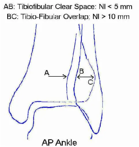
A. AP view
Identifies fractures of
malleoli
distal tibia/fibula
plafond
talar dome
body and lateral process of talus
calcaneous
Tib-fib Clear Space > 5mm or
Tib-fib Overlap < 10mm
Indicate syndesmotic injury
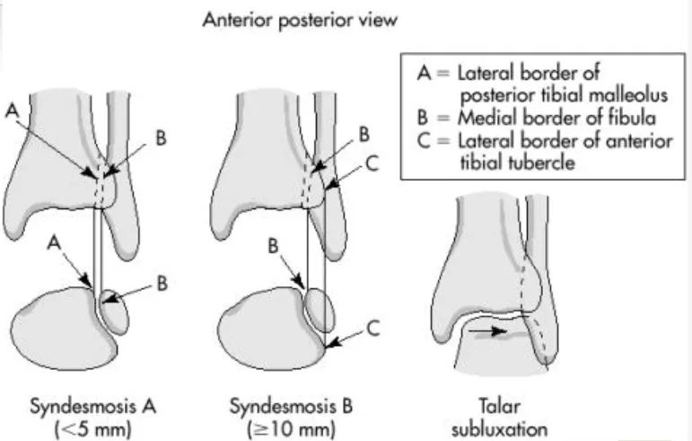
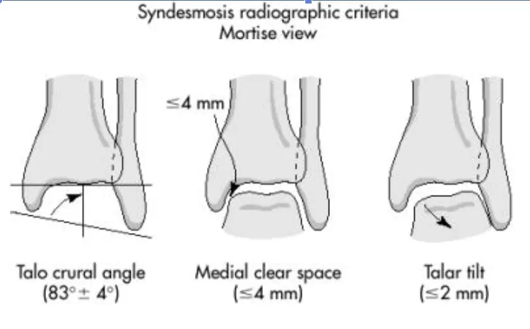
B. Mortise X-Ray
Taken with ankle in 15-25 degrees of internal rotation
Useful in evaluation of articular surface between talar dome and mortise
Tibio-fibula overlap about 1 mm
Tibiofibular clear space < 6 mm
Medical clear space < 4 mm
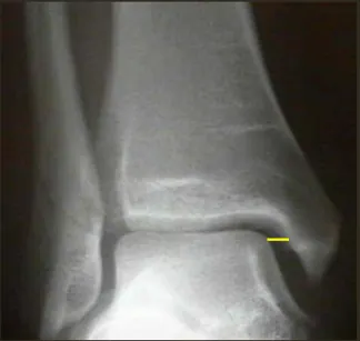
Medial clear space
Between lateral border of medial malleous and medial talus
<4mm is normal
4mm suggests lateral shift of talus
Medial clear space : lateral malleolar Fx. or Deltoid diosruption
C. Lateral X-ray
Identifies fractures of
Anterior/posterior tibial margins
Talus
Displacement of talus
Os trigonum
Anterior and posterior margins of distal tibia, base of 5th metatarsal bone
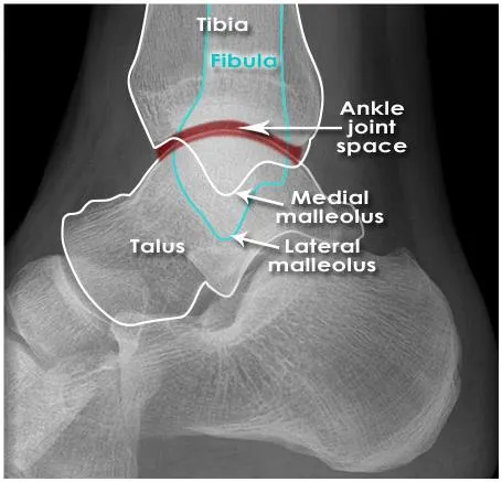
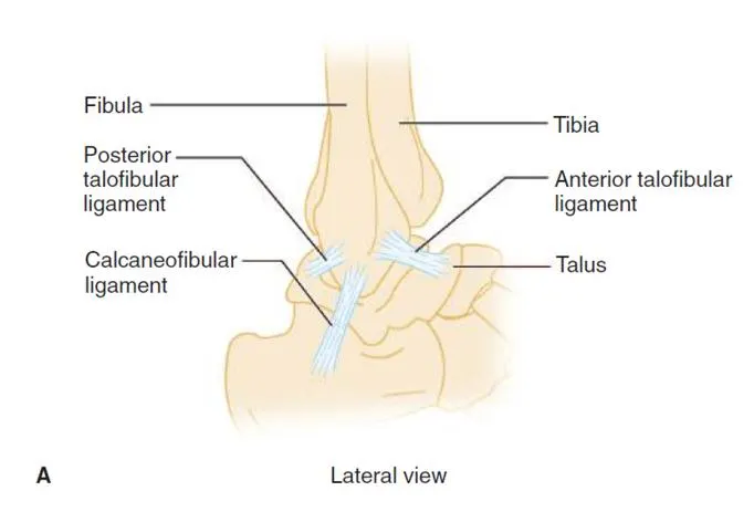
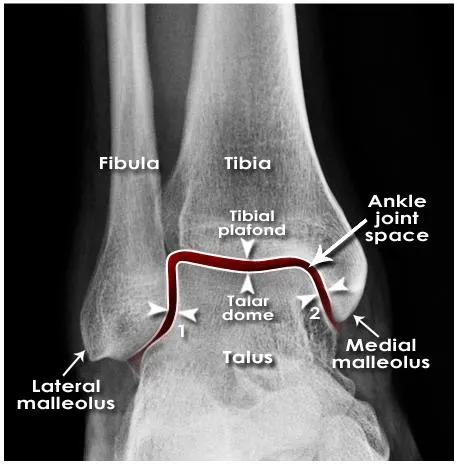
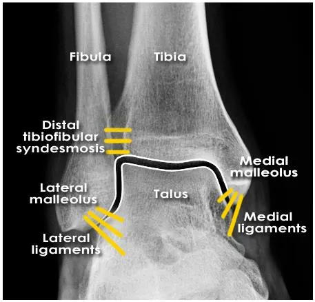
Ankle bone and lig. anatomy
Not visible
Joint space
understanding of the anatomical position of ligaments is required to appreciate the presence of ligament injuries
Ankle anatomy (Lig.)
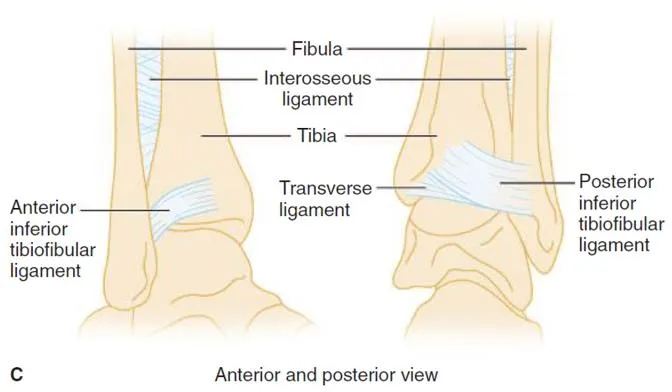
Syndesmotic ligament complex
Axial, rotational, & translational stability
Post. inf. Tibiofibular ligament 가 ant.보다 더 두껍고 강하다
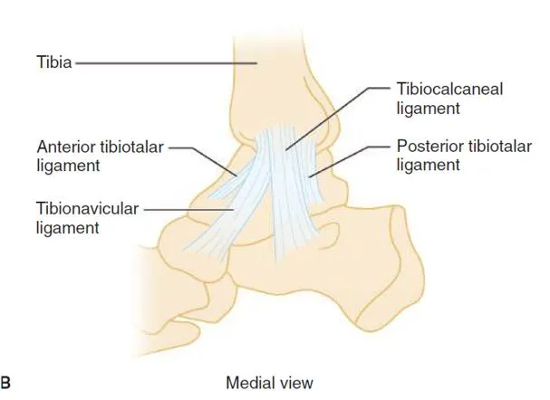
Deltoid (medial) ligament complex
가장 강한 인대 ⇒ eversion 보다 inversion이 자주 된다.
Lateral (fibular collateral) ligament complex