

•
abdominal viscera 가장 흔하게는 small bowel loops가 peritoneal or mesenteric aperture를 통해 복강이나 골반강 내의 compartment 로 돌출되는 현상
•
Orifices: Congenital / Acquired
•
life-threatening condition and a surgical emergency 일 수 있다.
•
bowel entrapment 가 acute intestinal obstruction 으로 빠르게 진행할 수 있고, 내버려 두면 strangulation and ischemia 로 진행할 수 있다.
A. Clinical signs and symptoms
•
Nonspecific!
•
M/C: Nausea, Vomiting, Abdominal pain, Abdominal distension
•
Ranging from no symptoms to symptoms of acute small bowel obstruction
B. Classification
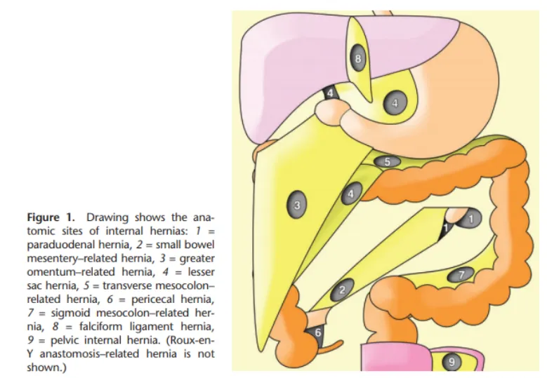
Orifice의 위치에 따라서 명명
예외 : Lesser sac, pelvic ineternal, Roux-en-Y anastomosis 관련 hernia는 다양한 orifice가 있을 수 있다.
가장 흔한 type은 para-duodenal hernia 임
C. Diagnosis
C1. Abdomen CT
(1) Bowel configuration
•
Sac-like mass or cluster of dilated small bowel loops가 정상적이지 않은 위치에 있는 상태
•
small bowel obstruction 양상
(2) Mesenteric abnormalities
•
vessels과 mesenteric fat이 hernia orifice로 수렴하는 모습
•
key mesenteric vessels의 주행이 displcement 되는 양상
•
Engorgement, crowding, twisting, stretching of mesenteric vessels if strangulation is present
(3) Position of surrounding viscera
•
Displacement of surrounding structures around the hernia sac
D. Para-Duodenal Hernia
•
Approximately 53% of all cases of internal hernias
•
Left-sided (75%) Right-sided (25%)
•
소장의 비정상적 회전 및 mesentery가 parietal peritoneum과 fusion되지 않아 발생
⇒ 소장의 loops 가 정상적이지 않은 peritoneal fossa로 들어가서 끼게 됨
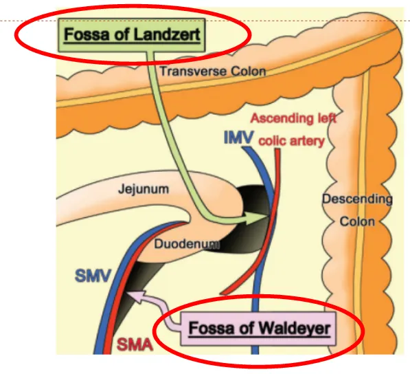
Duodenal segment나 jejunal loop가 Landzert’s fossa 라는 구멍으로 들어가서 끼임
Landzert’s fossa는 Duodenojejunal junction에 위치해 있고, 해부 시 약 2%에서 발견된다.
IMV와 Ascending Lt colic artery와 경계를 이루고 있다.
Bowel loop가 이 fossa로 들어가 끼이면 transverse mesocolon 좌측으로, 아래쪽으로 뻗어나가게 됨
RDPH는 Landmark를 SMA, SMV로 잡을 수 있음. 이 Waldeyer fossa는 선천적인 defect로 인구의 1%정도에서 발견됨.
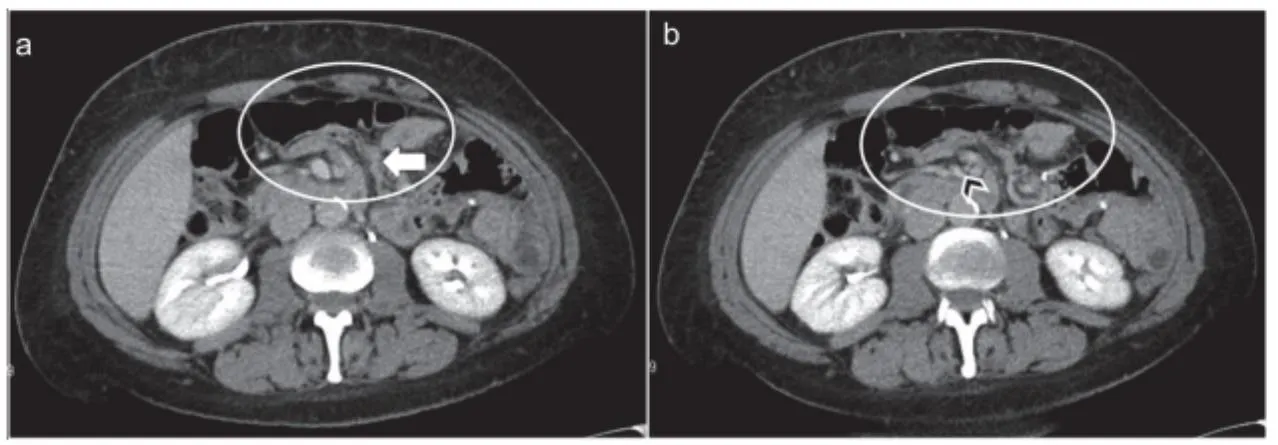
D1. Diagnosis
a: Bowel loop에 의한 mass effect로 stomach를 앞으로 밀어냄. encapsulated된 소장 보임
위와 췌장 사이에 위치할 수도 있다.
b: IMV와 Lt ascending colic a. 가 밀집되어 있고 확장되어 있다.
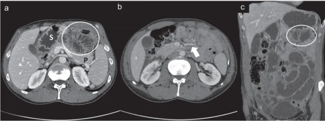
확장된 소장이 우측 복부에 위치 (상행결장과 십이지장의 3rd portion 보다 lateral에 위치)
c의 흰색 화살표: 확장되고 loop 로 수염되는 mesenteric vessel
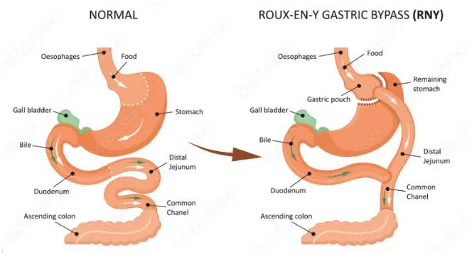
E. Roux-en-Y Anastomosis-related hernia
•
Incidence: 0.2% ~ 8%
•
Usually occur more than 1 month after surgery
•
Laparoscopic > Open approach
Lack of intra-abdominal adhesions
(prevent defects & fixation of the Roux-limb)
•
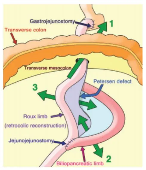
Trans-mesocolic : transmesocolon의 surgical defect로 돌출
•
Jejunostomy mesenteric : jejuno-jejunostomy site의 mesentery 로 돌출
•
Petersen type : Roux limb 뒤로 돌출. Roux limb의 mesentery와 transverse mesocolon사이 공간
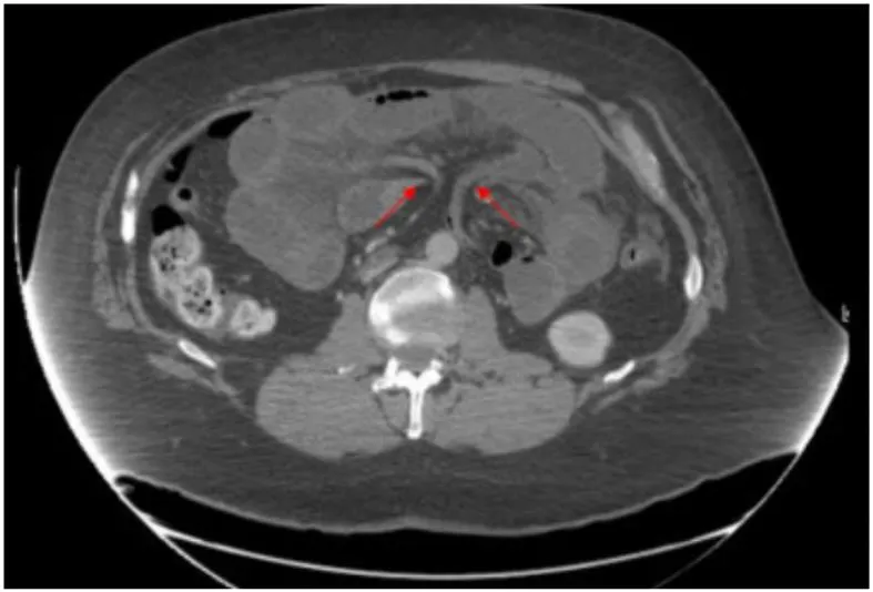
E1. CT Findings
(1) Bowel configuration
Small-bowel obstruction
(2) Mesenteric abnormalities
•
Swirled mesentery
•
Hurricane eye
: distal tubular mesentery with surrounding small loops
•
SMV beaking / Criss cross appearance
•
Mushroom sign : SMA와 distal mesentery vessel branch 사이 좁은 공간으로 mesenteric root가 지나가는데, 버섯처럼 보여서
•
Weeping mesentery (edematous mesentery with enlarged lymph nodes)
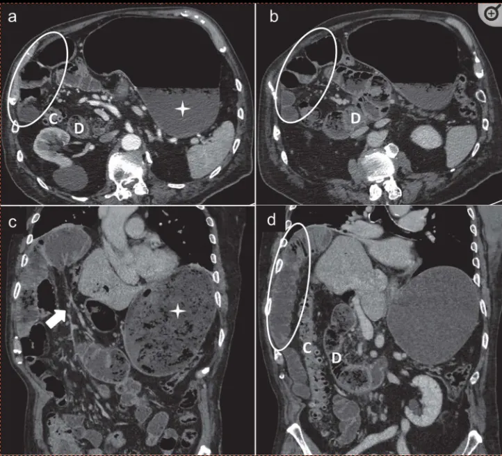
: Mesenteric swirl, small bowel obstruction 이 internal hernia진단에 있어 가장 정확도가 높다고
알려져 있으나, SMV의 직경이 새부리 모양처럼 줄어드는 SMV beaking sign 역시 흔하게 나타나는 sign이다.
a,b : 소장 loop가 복부 전벽에 인접하여 있다. SMV의 직경이 좁아져 beaked 모양으로 보인다 (화살촉)
herniated mesenteric loop 가 버섯 모양처럼 보인다.
(3) Surrounding viscera
Small-bowel behind superior mesenteric artery
Abnormal position of the jejuno-jejunostomy
