A. Anatomy
A1. Distal radius and ulna
(1) Distal radius
: Carpal bone과 직접 Articulation하는 유일한 forearm bone. (Fig 269-1)
(2) Distal ulna : Triangular fibrocartilage complex (TFCC) 에 의해 carpal bone과 분리
•
한 줄에 4개씩 2줄, 모두 8개
A2. Carpal bones
(1) Distal carpal row (trapezium, trapezoid, capitate, and hamate)
: 서로 붙어있고 metacarpal bone과 연결
(2) Proximal carpal row (scaphoid, lunate, triquetrum, and pisiform)
: Distal radius와 the distal carpal row 사이의 arch, "mobile link"
(3) 두 개의 palmar arches 사이 공간은 선천적으로 약한 구조로 이를 the space of Poirier 라고 함
B. Pathophysiology
•
손목 손상에서는 그 손상 기전을 이해하는 것이 도움이 된다.
•
대부분의 손상은 낙상하면서 팔을 쭉 뻗고 손목, 손이 dorsiflexion되면서 생긴다. (FOOSH)
•
Thenar area : Scaphoid와 그 주변 ligament들이 손상이 발생
•
Hypothenar area : Triquetrum, pisiform과 그 주변 ligament에서 손상이 발생
B1. 연령대 손상 ★
(1) Children
① Immature, weak한 radius의 epiphyseal plate, metaphysis에 잘 생김
② 상대적으로 cartilaginous한 carpal bone 손상은 적다.
(2) Young adults
: Scaphoid, proximal row intrinsic ligaments, distal radial metaphysis
(3) Elderly
① 골다공증 영향으로 여성에게 더 흔함
② Weak point는 distal radial metaphysis임
→ Colles fracture 유발 (often with intra-articulr involvement)
③ 또한 주로 fall로 인한 carpal bones의 injury가 높은 편
C. Clinical Features
Figure 269-3
C1. 양쪽 손목을 같이 관찰
C2. Anatomic snuffbox
(1) Wrist의 dorsum에서 가장 중요한 landmark
: 다음의 3가지가 만드는 triangle
① Bony radial styloid 의 proximal base
② The extensor pollicis brevis tendon 의 radial aspect
③ The extensor pollicis longus tendon 의 ulnar aspect
(2) Scaphoid가 이 triangle에서 만져질 수 있음. 만약 압통이 있으면 scaphoid 골절 의심 ★
C3. Lister's tubercle
(1) Distal radius 의 bony prominence 를 감싸는 EPL tendon.
(2) 여기의 바로 distal 부위가 scapholunate joint
만약 여기에 tenderness 있다면 scapholunate ligament injury 및 lunate fracture 의심.
C4. Scaphoid shift test
(1) Scapholunate ligament injury를 assess.
(2) 손목을 ulnar deviation 시킨 채 엄지로 scaphoid tuberosity를 누르면서 radial deviation 시킨다.
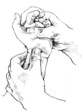
(3) 만약 ligament injury가 있다면 palpable "clunk"가 느껴짐
C5. Ulnocarpal stress test
(1) 손목을 ulnar deviation 시키고 눌렀을 때 pain이나 click이 있다면 triangular fibrocartilage complex injury를 시사한다.
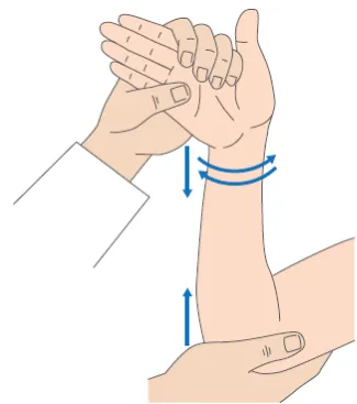
•
Ulnar styloid (Wirst의 ulnar aspect의 bony prominence_
: Triquetrum, triangular fibrocartilage complex가 distal portion 에 위치
: Ulnar styloid 위 압통이 있으면, styloid나 triangular fibrocartilage complex injury 의심
C6. Piano key sign
(1) Pronation 상태에서 forearm을 지탱하는 ulnar head가 눌려지면서 튕겨 나온다면, distal radioulnar joint injury를 시사한다. or TFCC 손상
D. Imaging
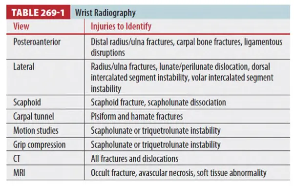
D1. Wrist의 Standard views
(1) Posteroanterior (PA)
(2) Lateral
(3) Oblique views
•
하지만 specific injury 시 다른 view 필요할 수도 있음
D2. Gilula lines을 관찰할 것
: Three smooth arcs outline the articular surfaces ar the radiocarpal and midcarpal joints
D3. Carpal bone은 서로 1~2mm 간격을 두고 퍼즐 모양으로 서로 맞춰져 있다.
D4. Scaphoid
(1) 정상적인 모습은 elongated shape
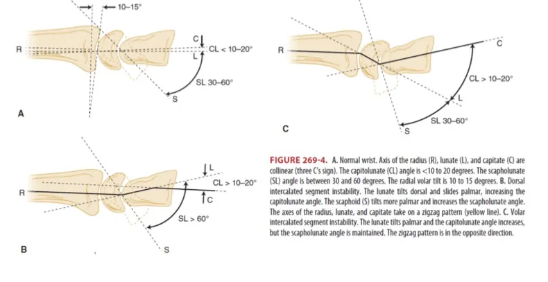
(2) Lateral view에서 정상적으로 palmar-flexed : lunate와 30~60°angle을 이룸 (Figure 269-4B & C).
(3) 만약 fracture나 lig. disruption이 있다면 보다 palmar rotation하게 되어, PA view에서 보면 정상 보다 짧아져 보인다.
(4) Scaphoid injury 시 scaphoid fat stripe를 obscure
D5. Radial styloid
(1) Distal radioulnar joint에서 8~18mm 정도 projection
(2) PA view에서 13~30°ulnar의 inclination을 만든다.
D6. Radius, lunate, capitate의 axis
: lateral view에서 보면 collinear 하다.
D7. Distal radius fx : m/c wrist fx
(1) Displaced fx는 deformity가 훨씬 뚜렷하지만, distal radial articular surface의 정상 volar tilt(10~15°)의 변화가 있는 경우가 손목 기능 예후에 더 큰 영향을 준다.
E. Ligamentous Injuries
•
Lunate : 손목의 중앙에 위치, Ligament injury도 주로 여기서
E1. Scapholunate Ligament Instability
(1) Wrist ligament injury 중 가장 흔함.
(2) 3가지 radiologic findings가 있다.
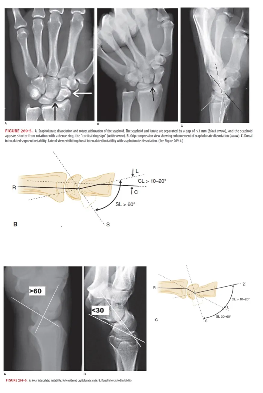
① Scapholunate dissociation
: Widening of the scapholunate joint space of >3 mm on the PA view (Figure 269-5A)
② Rotary subluxation of the scaphoid
: Scaphoid가 palmar 쪽으로 더 치우치고60°
(lateralview에서 scapholunate angle이 이상 증가 Figure 269-5B)
③ Dorsal intercalated segment instability (Figure 269-4B)
(3) ED treatment : radial gutter splint or short arm volar posterior mold
E2. Triquetrolunate Ligament Instability
(1) Lateral radiography : "volar intercalated segment instability" pattern (F269-4C, 269-6)
(2) ED treatment : ulnar gutter or short arm posterior mold
E3. Perilunate and Lunate Dislocations
* 10% of all carpal injuries
(1) Perilunate dislocation
① Lateral view에서 잘 보인다.
② Three C‘s sign(radius, lunate, capitate의 axis들이 거의 일직선을 이루는 것, normal sign) disruption 되고, capitate가 dorsal 쪽으로 displacemenet 된다.
③ Lunate는 radius가 계속 붙어 있다.
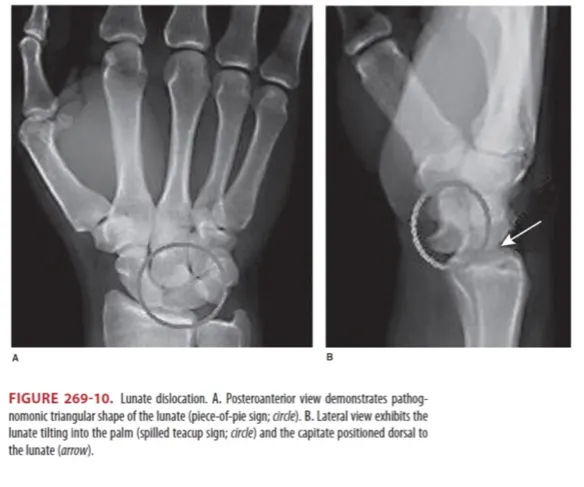
(2) Lunate dislocation
① Lunate가 radius에서 멀어지고 palm 쪽으로 위치하게 되는, ‘spilled teacup sign’이 보인다 (Figure 269-10B).
(3) Treatment
① Perilunate나 lunate dislocations 은 응급수술에 대한 협진필요
② CR & LAS 하거나 수술방 가서 ORIF 할 수 있다,
③ Complication
- Avascular necrosis : m/c cause 중 하나
- Early degenerative arthritis
- Delayed union
- Malunion
- Nonunion
- Avascular necrosis
- Median n. compression from the volar dislocation of the lunate into the carpal tunnel
F. Carpal Bone Fractures★
•
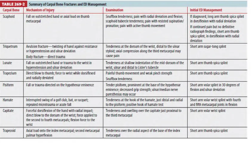
치료는 표 269-2를 일괄적으로 참조할 것 * 발생 빈도 순서대로 나열됨.
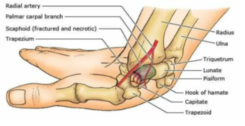
F1. Scaphoid Fracture
(1) M/C carpal bone fx.
(2) Anatomic snuffbox 에 localization 되고, wrist의 radial aspect 따라 통증 느낌
(3) proximal fx. area에 avascular necrosis가 생겨 disabiling arthritis로 진행될 수 있다.
(4) X-ray 상 10%에서는 확인을 못할 수 있음 → 임상적인 의심이 중요
(5) Non-displaced fx. : short arm thumb spica splint (임상적인 의심만 있을 시에도 splint 적용)
(6) Unstable fx : long arm thumb spica splint
•
Scaphoid bone은 distal 1/2 쪽으로 artery 가 들어와서 proximal 1/2 쪽으로
분지를 내는 방식으로 혈액공급. 따라서 weist fx.시 prox 1/2 의 무혈성 괴사 가능
F2. Triquetrum Fracture
(1) 2nd common carpal fx
(2) Dorsal avulsion fx. 시 lateral 및 oblique view에서 잘 보임 (”pooping duck”)
F3. Lunate fracture
(1) Blood supply가 distal end 쪽으로 들어오기 때문에, fracture 시 proximal쪽으로 avascular necrosis가 생길 수 있다.
F4. Trapezium fracture
(1) Displaced fracture 1mm 이상 및 diastases 2mm 이상이면 수술 필요
F5. Pisiform fracture
(1) Guyon's canal : Pisiform & hook of hamate로 이루어지며 ulnar n. & a.가 주행한다
F6. Hamate fracture
(1) Standard 및 carpal tunnel views 가 진단에 필요할 수 있다.
(2) Occult fx. 에서는 bone scan 이나 CT로 진단해야 할 때도 있다.
(3) P/Ex를 통한 Guyon’s canal injury d/dx 필요.
F7. Capitate Fracture
(1) Carpal bone에서 가장 큰 뼈로 대개 scaphoid fracture와 동반하여 생김. (Scaphocapitate syndrome)
(2) PA view에서 가장 잘 찾을 수 있다.
F8. Trapezoid fracture : 매우 드물다
G. Distal radius and ulnar fractures ★
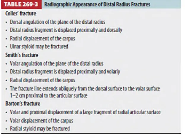
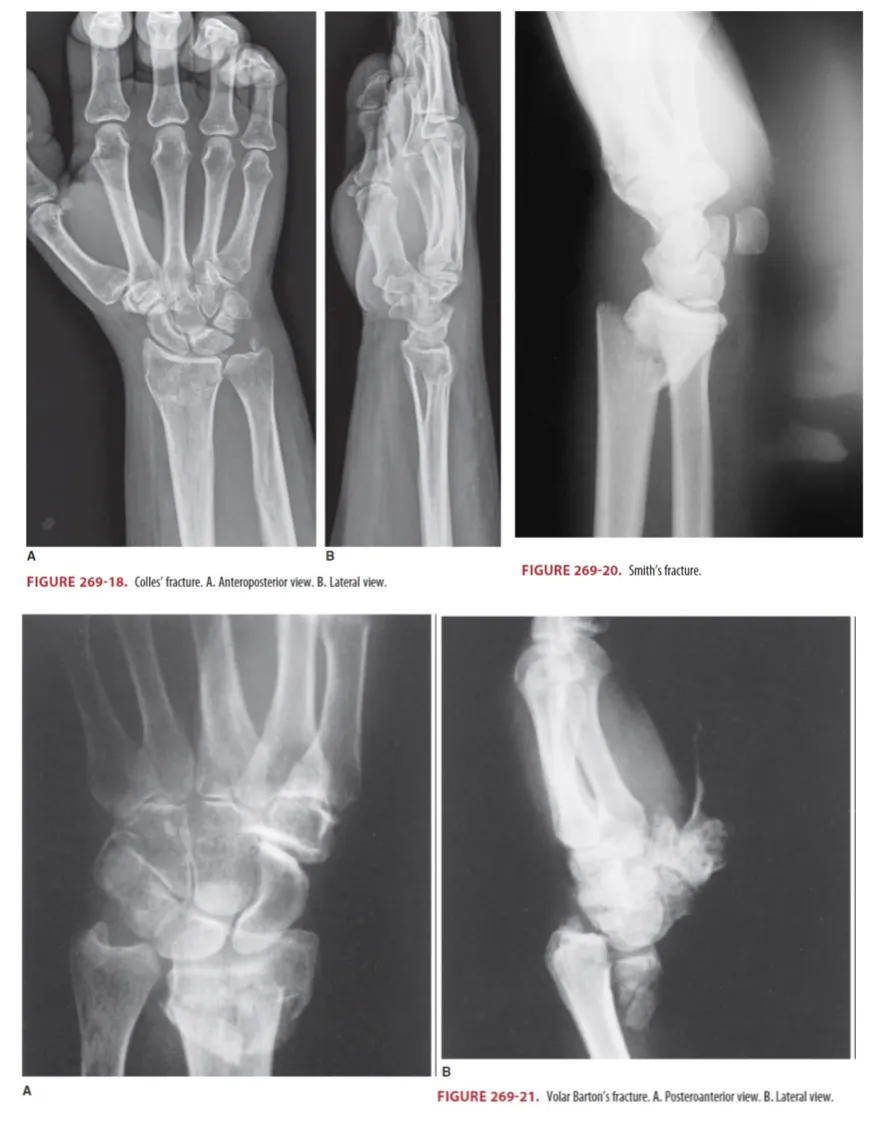
G1. Colle’s fracture ★
(1) 손을 밖으로 쭉 뻗은 상태에서 떨어져서 생기는 경우가 가장 흔함 (Figure 269-18)
(2) Ulnar styloid fx. 가 동반될 수 있는데 이는 TFCC 손상 동반 가능성 있음
(3) 특징적인 Wrist의 dorsiflexion, "dinner-fork" deformity 보임
(4) Dorsal angulation 및 comminution을 보는데 lateral view가 나음
(5) Unstable fx Angulation 20°,이상 intra –articular involvement comminute fx.
→ 대부분 closed reduction과 sugar tong splint로 치료될 수 있음
(6) Open fx. 이거나 신경-혈관손상 의심시는 즉각 OS evaluation
(7) Reduction 목표 : the volar tilt, radial inclination, a radius length의 정상화
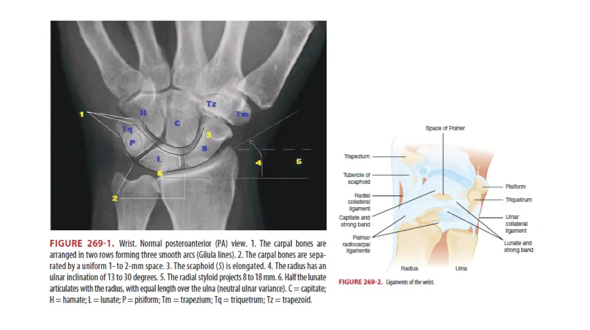
G2. Smith Fracture (Figure 269-20)
(1) Reverse colle’s fx. 로 불림. lateral view에서 volar angulated 되어 있음 (colle’s 와 반대)
(2) Lateral view가 angulated and displaced fx.를 가장 잘 보여준다.
(3) Garden-spade deformity
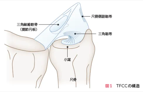
G3. Barton fracture (Figure 269-21)
(1) Distal radius의 dorsal 또는 volar rim이 골절되면서 때때로 Fx-DL이나 subluxation 을 만든다.
(2) Minimal displaced fx. 는 sugar tong splint apply & OS f/u
(3) Unstable fx.
: Radial articular surface를 50% 넘게 involve했거나, carpal subluxation이 동반되는 경우, ORIF 필요하므로 OS consult 시행함.
G4. Radial Styloid Fracture (Figure 269-22)
(1) 종종 Lunate dislocation 동반
(2) Treatment : Short arm splint
G5. Ulnar styloid fracture
(1) Forced radial deviation, dorsiflexion, or rotatory stress로 유발
(2) 단독으로로 발생가능하나, Colles Fx 등의 골절과 동반가능
(3) TFCC손상 동반가능
(2) Treatment : Ulnar gutter splint
G6. Distal Radioulnar Joint Disruption
(1) 보통 intra-articular fx. or distal radial shaft fx. 에서 보인다. (Galeazzi fracture-dislocation)
(2) Lateral radiograph
① Ulna의 volar or dorsal displacement가 나타난다.
② 정상적으로는 중앙에서 radius를 감싸는 모습