

•
흉통을 주소로 내원한 환자의 EKG에서 ST elevation이 관찰될 때, 15%만이 STEMI가 final diagnosis이다. 따라서 다음과 같은 상황들과 감별해야 한다.
•
ST elevation secondary to LVH
•
ST elevation secondary to conduction defect(LBBB, non-specific IVCD)
•
Early repolarization (Notched J-point in anterolateral leads)
•
Aneurysm
•
Old MI
•
Pericarditis/Myocarditis
•
WPW syndrome
•
Brugada pattern
•
Takosubo syndrome
•
Hyperkalemia, Hypercalcemia
흉통을 호소하는 환자의 심전도에서 ST elevation이 관찰될 때 약15%만이 최종적으로 STEMI로 진단되며 나머지는 심전도상에서 ST elevation이 나타나는 다른 질환들로 최종 진단되며 이러한 현상을STEMI mimics라고 합니다. STEMI mimics을 보일 수 있는 질환들로는 LVH, LBBB, Early repolarization, Pericarditis 등이 있습니다.
참고로 이러한 원인 질환들을 외우기 위한 mnemonic이 있는데 “elevation”으로 외우시면 됩니다.
Electrolytes (Hyperkalemia)
Left Bundle Branch Block
Early Repolarization
Ventricular Hypertrophy (Left)
Aneurysm (Ventricular)
Thailand (Brugada Syndrome)
Inflammation (Pericarditis)
Osborne (J) Waves (hypothermia)
Non-Ischemic Vasospasm
<ST elevation에서 acute STEMI를 더 시사하는 소견>
1.
이전 EKG와 비교하여 새로운 ST segment의 변화 & dynamic change
2.
Reciprocal ST depression
Ratio of T wave to QRS complex amplitude
•
T wave/QRS ratio < 0.36 in all precordial leads : LV aneurysm를 더 시사함
•
T wave/QRS ratio > 0.36 in any precordial leads : Anterior STEMI를 더 시사함
가. Conduction defect에 의한 STE (LBBB)
<Sgarbossa's criteria>
•
Criteria A : Concordant STE >1mm in leads with a positive QRS complex (score 5)
•
Criteria B : Concordant STE >1mm in V1-V3 (score 3)
•
Criteria C : Excessively discordant STE >5mm in lead with negative QRS complex (score 2)
<Smith's modification>
•
≥ 25% of the depth of the preceding S-wave.
•
ST elevation이 관찰될 시, Sgarbossa's criteria를 적용할 경우 3점 이상에서 MI에 대한 specificity가 98%로 높으나 sensitivity는 20%로 낮다.
•
Smith modification을 적용하면, specificity도 높고, sensitivity도 80%까지 증가시킬 수 있다.
•
Sgarbossa's criteria는 결국 Acute MI가 없는 LBBB에서는 ST segment와 T wave가 QRS의 방향과 discordant 로 나타난다는 것이다.
나. True STEMI vs LVH with strain
•
LVH는 depolarization, repolariation의 change를 유발한다.
•
이는 ECG상의 QRS duration widening, increase QRS amplitude, changes in the ST segment and T wave를 일으킬 수 있다.
나.1. Typical한 LVH strain
•
STD & T wave inversion in lateral leads
•
STE in leads V1-3 (discordant to deep S wave)
•
하지만, 다른 형태의 strain 또한 나타날 수 있으며 이는 LVH 환자들이 STEMI로 false positive하게 진단되는 경우가 있다.
•
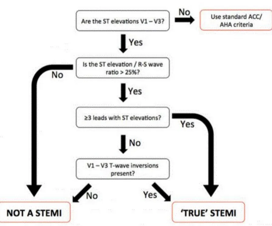
이의 개선을 위하여 2014년에 이뤄진 cohort연구의 결과에 따르면(Reference 1), patients with ECG LVH를 대상으로 STEMI 진단에 기존 ST elevation의 threshold를 정한 ACC/AHA criteria를 적용할 경우, sensitivity는 73%, specificity는 58%였다. Cohort data를 통해 얻어낸 criteria (사진1)을 적용 할 경우, sensitivity는 77%, specificity는 91%로 기존의 criteria에 비해 상승하였다. 해당 연구에 따르면, St segment elevation to R-S wave magnitude의 ratio가 LVH와 true stemi의 구분에 있어서 중요하다는 점을 새롭게 제시한다.
Reference)
•
Ehrin J. Armstrong, Ameya R. Kulkarni, Prashant D. Bhave. Electrocardiographic Criteria for ST-Elevation Myocardial Infarction in Patients With Left Ventricular Hypertrophy. The American Journal of Cardiology, Volume 110, Issue 7, 2012, Pages 977-983,
•
Edhouse J, Thakur RK, Khalil JM. ABC of clinical electrocardiography. Conditions affecting the left side of the heart. BMJ. 2002 May 25;324(7348):1264-7
•
Yochai Birnbaum, Mahboob Alam. LVH and the diagnosis of STEMI - how should we apply the current guidelines?. Journal of Electrocardiology. Volume 47, Issue 5. 2014,
다. True STEMI vs LV aneurysm
•
Acute STEMI 이후, ST segment는 통상 2주간 baseline으로 돌아온다.
•
하지만 anterior STEMI 60%의 환자, inferior STEMI 5%의 환자에서 ST elevation이 지속되고, 이러한 기전은 acute MI 이후 incomplete reperfusion 과 transmural scar formation 때문이다.
•
이러한 EKG 패턴은 ventricular wall의 paradoxical movement와 관계되어있다.
나.1. STE에서 LV aneurysm을 시사하는 소견>
1.
acute MI 이후, ST elevation이 2주 이상 지속
2.
Precordial lead에 주로 보인다.
3.
Concave하거나 convex한 양상을 보인다.
4.
Well formed Q wave
5.
Abscence of dynamic ST segment change & reciprocal ST depression
6.
T wave는 QRS complex에 비해 small amplitude를 보인다.
라. True STEMI vs Early repolarization
•
BER에서 STE를 관찰할 수 있으나, 이는 젊고 건강한 사람에서 나타날 수 있는 정상 소견으로 STEMI와의 감별을 요구한다.
라.1. Early repolariazation ECG findings
1.
precordial lead, Lead 2,3에서 ST elevation 보이며 aVR depression 보임
2.
Pathologic Q가 없는 것이 특징
라.2. BER에서 STEMI를 더 시사하는 소견
1.
>5mm 의 ST elevation
2.
Non-concave ST elevation
3.
Inferior reciprocal changes
4.
Anterior ST depression
5.
Terminal QRS distortion in V2 or V3
6.
V2, V4에 Q wave가 보일 때
7.
V2~V6 에 T wave inversion이 보일 때
E. Acute pericarditis
F. Brugada syndrome
Brugada’s Syndrome은 남자에 흔히 나타나고 a prominent J-wave with a characteristic
downsloping ST-segment elevation in leads V1–3
Brugada’ syndrome은 동남아에 흔하고 (sudden unexplained nocturnal death syndrome),
sudden cardiac death에 위험성이 높기 때문에 잘 알아야하고 internal cardioverter-defibrillator placement로 예방할 수 있다.
Brugada synd.은 우성유전으로 발현되며, 그 결과 sodium channel 기능의 완전 소실 또는
sodium channel 활성화 후 회복의 가속으로 나타난다.
