


1.
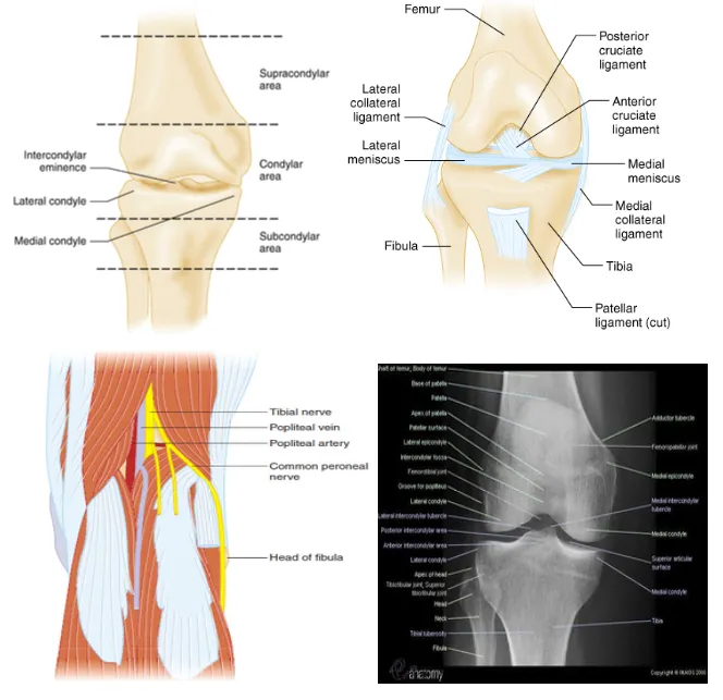
Four ligament of knee : ACL, PCL, MCL, LCL

1.
Popliteal fossa : Popliteal artery 公 vein, common peroneal nerve, tibial nerve
2.
무릎 신체검진은 크게 5가지 구성요소,즉 병력,관찰(observation), 시진,촉진,부하검사(stress test)로 이루어진다.
3.
보행,관절의 functional ROM, 약간의 저항을 줄 때 굽혀진 무릎을 필 수 있는지 확인하고,붓기,반상 출혈 (ecchymosis), 삼출 (effusion), 종괴,슬개골의 위치와 크기,근육의 부피,발적,국소 손상의 증거를 시진한다.
4.
Supine position에서 다리 길이가 동일한지 확인하고 환자에게 best possible active ROM을 보여 달라고 요청한다.
5.
Pulse와 distal neurologic function을 평가한다.
6.
모든 검진에서 정상 무릎과 비정상 무릎을 비교해야 하며 특히 촉진과 부하검사에서 중요하다. : 무릎을 촉진할 때는 아프지 않은 곳부터 시행하여 환자의 지각을 최소화하고 patellar patellar
facet들과 femoral, tibial condyle을 촉진하여 통증과 crepitus를 확인 후 관절의 삼출 압통, 열감,근력 감각, 박동의 위치를 기술한다.
A. Anatomy
•
Tintinalli 9th, p1850, Fig 274-1, 2, 3
•
Four ligament of knee : ACL, PCL, MCL, LCL
•
Popliteal fossa : Popliteal artery & vein, common peroneal nerve, tibial nerve
B. Clinical feature
B1. Neurovascular injury
(1) Popliteal artery injury
•
주요 원인 손상
◦
femoral condyle fracture
◦
displaced tibial plateau fracture
◦
isolated PCL injury
◦
multiple ligament injury
◦
knee dislocation
•
Collateral circulation 부족하여 8시간 안에 회복되지 못하면 amputation!
⇒ 건측과 distal pulse 비교 (민감도 79%, 특이도 91%), ABI, duplex US, angiography
•
Compartment syndrome, venous injury, arterial thrombosis 발생 주의
(2) Peroneal nerve injury
•
Severe ligament injury, dislocation 시 발생
•
fibular head fracture or avulsion의 절반 가량에서 발생
•
Deep peroneal nerve
: 첫째-둘째 발가락 사이 dorsal web space sensation, foot dorsiflexion, toes extension
→ Injury 발생 시 foot drop, gait difficulty
C. Diagnosis
C1. Physical Exam
•
무릎 신체검진은 크게 5가지 구성요소, 즉 병력, 관찰(observation), 시진, 촉진, 부하검사(stress test)로 이루어진다.
•
보행, 관절의 functional ROM, 약간의 저항을 줄 때 굽혀진 무릎을 필 수 있는지 확인하고, 붓기, 반상 출혈 (ecchymosis), 삼출 (effusion), 종괴, 슬개골의 위치와 크기, 근육의 부피, 발적, 국소 손상의 증거를 시진한다.
•
Supine position에서 다리 길이가 동일한지 확인하고 환자에게 best possible active ROM을 보여 달라고 요청한다.
•
Pulse와 distal neurologic function을 평가한다.
•
모든 검진에서 정상 무릎과 비정상 무릎을 비교해야 하며 특히 촉진과 부하검사에서 중요하다.
•
무릎을 촉진할 때는 아프지 않은 곳부터 시행하여 환자의 지각을 최소화하고 patellar와 patellar facet들과 femoral, tibial condyle을 촉진하여 통증과 crepitus를 확인 후 관절의 삼출, 압통, 열감, 근력, 감각, 박동의 위치를 기술한다.
•
무릎을 flexion한 상태에서 patellar의 크기, 모양, 위치를 검진하고
•
무릎을 extension 시키며 patellar의 유동성을 확인한다.
C1. Imaging
(1) Ottawa knee rules
•
Fracture 확인에 sensitive하며 응급실에서의 대기시간, 비용을 절감

(2) Pittsburgh knee rules (Fig 274-4)
•
Ottawa Rules보다 specificity가 좋다. 두 rules 모두 소아 >2세, 성인에게 적용

(3) X-ray
•
A-P, Lat. view가 기본
•
Lateral view
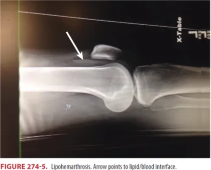
◦
X-ray 상 보이는 fat fluid level (lipohemarthrosis)는 intra-articular fracture 의미(white arrow)
•
Oblique views
◦
특히 미세한 tibial plateau fracture를 발견하는데 유용
◦
Internal oblique view는 lateral plateau 평가에 좋다
◦
external oblique view는 medial plateau 평가에 좋다
•
Tunnel or intercondylar view
◦
Intercondylar region, 특히 tibial spine fracture를 확인
•
Sunrise (skyline, axial, or tangential) view
◦
Conventional views 에서 놓칠 수 있는 patella의 non-displaced vertical or marginal fracture 보는데 용이
◦
Patellar subluxation, fracture 의심 시 사용
D. Specific injury
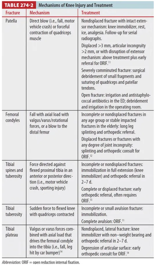
D1. Patella fractures

(1) Transverse fractures가 가장 흔하고, stellate, comminuted fracture 순
: Transverse fracture는 displacement 가능성이 높으며 disrupted extensor mechanism과 연관
(2) Extensor mechanism intact(중력에 대해 다리 펴고 들어올리기 가능)하고, 2mm 미만 step off,
3mm 미만의 fracture displacement 시 비수술적 치료
D2. Femoral condyle fractures
: 신경과 혈관 손상 - 흔하지 않지만 체크
•
Popliteal artery 손상 가능성 : distal pulse
•
deep peroneal nerve 첫번째와 두번째 발가락 사이(dorsal web space) 감각
D3. Tibial spine & tuberosity fractures
(1) Tibial spine 단독 손상은 드물며, ant. 가 post.에 비해 10배 흔함
→ Cruciate ligament insufficiency
(2) Hemarthrosis, full extension 불가, Lachman test 양성
D4. Tibial plateaus fractures
(1) Lateral plateau fracture : Medial에 비해 흔하며 ACL, MCL 손상과 연관
(2) Medial plateau fracture : PCL, LCL 손상과 연관
(3) Segond`s fracture : ACL 손상에 특징적 (뒤에 다시)
(4) 합병증 : Popliteal artery injury, DVT, OA
E. Ligamentous and Meniscal injuries
•
Hyperextension, valgus & varus stresses, A-P displacement에 의해 유발
•
Valgus stress에 의한 손상이 훨씬 흔하며 knee의 medial side injury 유발
•
Varus stress는 lateral side injury
•
MCL, LCL, ACL, PCL, capsular structure, medial & lateral meniscus 손상 가능
E1. Medial & lateral collateral ligament injury

(1) MCL → Valgus stress test로 확인
(2) LCL → Varus stress test로 확인
(3) Valgus, varus test (30도 flexion)에서 시행 시
•
이완(laxity)이 제한되며 pain 유발 시 strain 가능성이 높음
•
정상 무릎과 비교 시, 명확한 end point 없이 이완되면 medial or lateral collateral lig.의 complete rupture 의심
(4) Full extension에서 시행 시
•
valgus laxity ⇒ MCL complex 전체, cruciate lig., post. capsule tear 포함한 심각한 손상 의미
•
varus laxity ⇒ knee의 posterolateral corner, cruciate ligament를 포함한 심각한 손상 및 peroneal nerve 손상 가능성을 의미
E2. Anterior cruciate ligament injury
•
Tibia의 deceleration, hyperextension, 과도한 internal rotation에 의해 유발
•
손상 시 발생하는 “pop(popping noise)”이 특징적
•
수 시간 안에 swelling, instability가 발생
•
traumatic effusion 또한 ACL disruption을 의심할만한 소견
(1) Lachman test
•
Sensitivity 및 specificity가 가장 높다 (84%, 100%).
•
30도 정도 무릎을 flexion 시킨 상태에서 한 팔로 thigh 고정하고
lower leg를 앞으로 당겼을 때,
⇒ 반대쪽 무릎과 비교하여 displacement 있을 시 ACL injury 의심

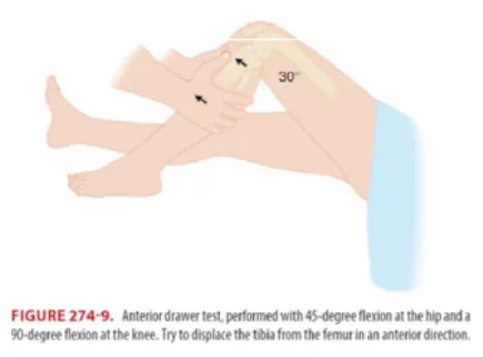
(2) Anterior drawer test
•
오랜 기간 사용되어왔던 방법이나, sensitivity는 62% 정도
•
Hip을 45도 flexion, knee를 90도 flexion시킨 다음 tibia를 femur로부터 ant. 쪽으로 displacement 시켰을 때
⇒ 반대쪽 무릎에 비해 6mm 이상 displacement 될 시 ACL injury 의심
(3) Pivot shift test
•
Heel을 들어 올리면서 hip을 45도 flexion, knee를 full extension 시킨 다음, 한쪽 팔로 knee, fibular head를 잡고 ankle이랑 knee를 internal rotation하면서 knee flexion 시켰을 시,
⇒ 20~30도 가량에서 딸깍하는 느낌이 들면 양성
•
Anesthesia 없이 시행 시 sensitivity 24%, specificity 98%

E3. Posterior cruciate ligament injury
(1) 단독 손상은 흔하지 않으며 post. drawer test, sag sign을 통해 확인
* Posterior drawer test : Ant. 와 같은 자세에서 post.로 displacement 발생 시 의심
•
Sag sign
: PCL 손상 시 hip 45도, knee 90도 flexion 자세에서 중력에 의해 tibia가 후방으로 내려와 sag 모양
or tibia tubercle의 drop back 발생

F. Hemarthorosis or effusion
•
Hemarthrosis가 있으면 knee ligament injury를 의심해 볼 수 있으며, 가장 흔한 것은 ACL 손상.
: 오히려 심한 ligamenet injury의 경우에는 완전히 파열되어 blood가 조직 내로 빠져나가 pain 별로 없고 hemathorosis 없을 수 있음
•
Traumatic hemarthrosis는 보통 손상 수분~수시간 내에 발생하고 chronic effusion은 1~2일에 걸쳐 synovial inflammation에 의해 유발
•
Segond`s fracture
: Lateral tibial condyle의 lateral capsular ligament attachment site에서 avulsion fracture
→ ACL rupture marker
•
Medial tibial plateau에서 cortical avulsion(매우 드뭄)
→ PCL & medial mediscus tear
G. Treatment
G1. Ligamentous injury
(1) Single ligament injury (strain, rupture)
•
Knee immobilizer, ice packs, elevation, NSAIDs, and ambulation (가능한 정도만)
→ Knee immobilizer 사용 시 mobility를 유지, 구축 방지 위해 매일 ROM exercise 교육
→ ROM exercise는 ice pack apply 후 10~20도 flexion-extension, 하루 3~4차례
(2) Arthrocentesis
•
Effusion의 외상성 판단에 대한 진단 목적으로 시행 가능
•
치료 목적으로는 large, tense effusion인 경우 제외하고 evidence 부족
→ Aspiration 시 발견된 혈액과 지방입자는 lipohemarthrosis의 특징적인 소견이며 intra-articular
knee fracture를 시사
G2. Meniscal injury
•
ACL injury 가 흔히 동반되며 medial meniscus injury 가 lateral에 비해 2배 흔함
•
Joint line tenderness (민감도 70%, 특이도 15%)
•
McMurray test (약 50%대), grind test
•
P/E의 민감도 특이도가 낮아
문진 상 knee extension or flexion 시 painful locking 발생 여부가 진단에 도움
→ 확진은 MRI or arthroscopy
(1) McMurray test
•
앙와위에서 슬관절의 최대 굴곡 상태로 족관절부 또는 발꿈치를 잡고 회전시키면서 천천히 신전
•
신전 시 통증과 마찰음이 나오면 양성
•
완전 굴곡에서 90도 굴곡 사이 증상 → Meniscus post. horn의 손상 시사
•
그 후의 증상 → 반월판 연골의 중간부 또는 ant. horn손상 시사
•
외회전으로 내측 손상, 내회전으로 외측 손상을 진단
(손상 양상에 따라 회전의 방향과 일치하지 않게 나타날 수 있다.)

G3. Locked knee
•
심한 통증과 함께 actively or passively full extension 안되는 상태
(1) 원인
•
가장 흔한 원인은 meniscus tear
•
ACL rupture, patella dislocation, loose body, foreign body와 감별 필요함
(2) 치료
•
sedation 하 closed reduction
→ knee 90° flexion 상태로 다리를 들어올린 후 longitudinal traction, internal & external rotation
→ 실패 시 operative arthroscopy
G4. Knee dislocation
(1) Ant. dislocation이 m/c (약 40%), post. (33%), lat., med., rotary 순
(2) Knee dislocation은 severe injury가 있는 경우 spontaneous reduction이 약 50%에서 일어나므로
주의를 요하며 popliteal artery 또는 peroneal nerve injury (mostly with posterolateral dislocations), ligamentous & meniscal injury의 합병증 발생율이 높음
→ 특히 popliteal artery 손상 (환자의 1/3) 발생율이 높고 vascular reconstruction 지연 시 예후가 불량하므로 knee dislocation 모든 환자에게 arteriography 시행을 권유하기도
(3) Longitudinal traction 통해 reduction 시행, 20도 knee flexion 상태로 splint apply
→ Reduction 후 반복적인 neurovascular exam, ABI
→ Asymmetrical distal pulse or ABI<0.9 시 CT angiogram or ateriography,
reduction 전 pulse 없다가 reduction 후 돌아온 경우 ABI 시행 및 vascular surgery consult
(4) Knee dislocation 환자에서 normal distal pulse라도 popliteal artery injury 배제 불가
G5. Patellar dislocation
(1) 주로 lateral condyle의 lateral로 displacement, pain, deformity 발생
(2) Reduction
•
Conscious sedation 하에 hip flexion, knee hyperextension 상태에서 patella 밀어넣기
(3) Irreducible dislocation
•
Older age, preexisting patellofemoral arthritis, flexion <45도, anterolateral patellar position, patellar axis의 internal rotation → 수술
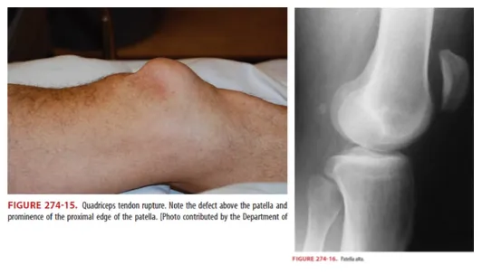
G6. Quadriceps or patellar tendon rupture
(1) 호발연령
•
Quadriceps tendon rupture 는 >40세 에서 주로 발생
•
Patellar tendon rupture 는 <40세 에서 주로 발생
(2) Tendinitis 과거력, steroid 사용이 risk factor
(3) Patella proximal or distal에서 tendon rupture에 따라 defect가 만져짐
(4) Patella alta (Figure 274-14) : Lat. x-ray에서 high-riding patella 소견
G7. Patellar tendinitis
(1) Jumper`s knee : runner, basketball & volleyball player
(2) NSAIDs, eccentric quadriceps-strengthening exercise, activity modification
→ Steroid inj. 금기
G8. Postarthroscopy problem
: Effusion이 m/c, infection은 매우 드물다.
