
Overview
최신 스캐너에서 사용할 수있는 많은 시퀀스를 생각하는 가장 간단한 방법은 조직 모양에 대한 지배적 인 영향에 따라 분류하는 것입니다.
이것은 모든 시퀀스를 proton density (PD) weighted, T1 weighted, T2 weighted, diffusion weighted, flow sensitive 및 '기타'로 나누어줍니다.
fat 또는 fluid attenuation (감쇠) 또는 contrast enhanced와 같은 여러 '선택 추가 기능'도 고려할 수 있습니다. 이는 다음과 같이 광범위한 범주로 이어집니다.
•
T1
◦
gadolinium enhanced
◦
fat suppressed
•
T2
◦
fat suppressed
◦
fluid attenuated
◦
susceptibility sensitive
•
proton density
◦
fat suppressed
•
diffusion weighted
•
flow sensitive
◦
MR angiography
◦
MR venography
◦
CSF flow studies
•
miscellaneous
◦
MR cholangiopancreatography (MRCP)
▪
a special T2-weighted sequence
◦
MR spectroscopy
◦
MR perfusion
◦
functional MRI
◦
tractography
Terminology
Intensity
음영의 강도에 따라 Intensity 를 다음과 같이 분류한다.
•
high signal intensity = white
•
intermediate signal intensity = grey
•
low signal intensity = black
Often we refer to the appearance by relative terms:
•
hyperintense = brighter than the thing we are comparing it to
•
isointense = same brightness as the thing we are comparing it to
•
hypointense = darker than the thing we are comparing it to
NB: the word density is for CT, and there are few better ways to show yourself as an MRI noob than by making this mistake.
Diffusion
When describing diffusion weighted sequences, we also use the term intensity but additionally we use the words restricted diffusion and facilitated diffusion to denote whether water can move around less easily (restricted) or more easily (facilitated) than expected for that tissue. Again many use these words as if they are absolute terms and this leads to confusion (more on this issue here).
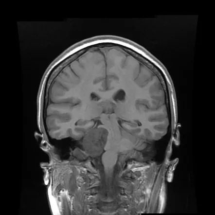
T1 weighted sequences
T1 weighted sequences are part of almost all MRI protocols and are best thought of as the most 'anatomical' of images, resulting in images that most closely approximate the appearances of tissues macroscopically, although even this is a gross simplification.
The dominant signal intensities of different tissues are:
•
fluid (e.g. urine, CSF): low signal intensity (black)
•
muscle: intermediate signal intensity (grey)
•
fat: high signal intensity (white)
•
brain
◦
grey matter: intermediate signal intensity (grey)
◦
white matter: hyperintense compared to grey matter (white-ish)
Contrast enhanced
The most commonly used contrast agents in MRI are gadolinium based. At the concentrations used, these agents have the effect of causing T1 signal to be increased (this is sometimes confusingly referred to as T1 shortening). The contrast is injected intravenously (typically 5-15 mL) and scans are obtained a few minutes after administration. Pathological tissues (tumours, areas of inflammation / infection) will demonstrate accumulation of contrast (mostly due to leaky blood vessels) and therefore appear as brighter than surrounding tissue. Often post contrast T1 sequences are also fat suppressed (see below) to make this easier to appreciate.
Fat suppression
Fat suppression (or attenuation or saturated) is a tweak performed on many T1 weighted sequences, to suppress the bright signal from fat. This is performed most commonly in two scenarios:
Firstly, and most commonly, after the administration of gadolinium contrast. This has the advantage of making enhancing tissue easier to appreciate.
Secondly, if you think that some particular tissue is fatty and want to prove it, showing that it becomes dark on fat suppressed sequences is handy.
T2 weighted sequences
T2 weighted sequences are part of almost all MRI protocols. Without modification the dominant signal intensities of different tissues are:
•
fluid (e.g. urine, CSF): high signal intensity (white)
•
muscle: intermediate signal intensity (grey)
•
fat: high signal intensity (white)
•
brain
◦
grey matter: intermediate signal intensity (grey)
◦
white matter: hypointense compared to grey matter (dark-ish)
Fat suppressed
In many instances one wants to detect oedema in soft tissues which often have significant components of fat. As such suppressing the signal from fat allows fluid, which is of high signal, to stand out. This can be achieved in a number of ways (e.g. chemical fat saturation or STIR) but the end result is the same.
Fluid attenuated
Similarly in the brain, we often want to detect parenchymal oedema without the glaring high signal from CSF. To do this we suppress CSF. This sequence is called FLAIR. Importantly, at first glance FLAIR images appear similar to T1 (CSF is dark). The best way to tell the two apart is to look at the grey-white matter. T1 sequences will have grey matter being darker than white matter. T2 weighted sequences, whether fluid attenuated or not, will have white matter being darker than grey matter.
마찬가지로 뇌에서 우리는 CSF의 눈부신 높은 신호없이 실질 체부를 발견하기를 원합니다. 이를 위해 CSF를 억제합니다. 이 시퀀스를 FLAIR이라고합니다. 중요하게도 언뜻보기에는 FLAIR 이미지가 T1과 유사하게 나타납니다 (CSF는 어둡습니다). 두 가지를 구분하는 가장 좋은 방법은 회색 - 흰색 물질을 보는 것입니다. T1 염기 서열은 회색 물질이 백색 물질보다 더 어둡게됩니다. T2 가중치 시퀀스는 유체 감쇠 유무에 관계없이 회색 물질보다 어두운 백색 물질을 갖게됩니다.
Susceptibility sensitive sequences
Being able to detect blood products or calcium is important in many pathological processes. MRI offers a number of techniques that are sensitive to these sort of compounds. Generally these sequences exploit what is referred to as T2* (T2 star) which is highly sensitive to small perturbations in the local magnetic field. The most sensitive of these sequences is known as susceptibility weighted imaging (SWI) and is also able to distinguish calcium from blood.
PD weighted sequences
Given that nuclear magnetic resonance of protons (hydrogen ions) forms the major basis of MRI, it is not surprising that signal can be weighted to reflect the actual density of protons; an intermediate sequence sharing some features of both T1 and T2.
Proton density images were extensively used for brain imaging, however they have largely been replaced by FLAIR. PD however continues to offer excellent signal distinction between fluid, hyaline cartilage and fibrocartilage makes this sequence ideal in the assessment of joints.
The dominant signal intensities of different tissues are:
•
fluid (e.g. joint fluid, CSF): high signal intensity (white)
•
muscle: intermediate signal intensity (grey)
•
fat: high signal intensity (white)
•
hyaline cartilage: intermediate signal intensity (grey)
•
fibrocartilage: low signal intensity (black)
Diffusion weighted sequences
Diffusion weighted imaging assess the ease with which water molecules move around within a tissue (mostly representing fluid within the extracellular space) and give insights into cellularity (e.g. tumours), cell swelling (e.g. ischaemia) and oedema.
The dominant signal intensities of different tissues are:
•
fluid (e.g. urine, CSF): no restriction to diffusion
•
soft tissues (muscle, solid organs, brain): intermediate diffusion
•
fat: little signal due to paucity of water
Typically you will find three sets of images when diffusion weighted imaging is performed: DWI, ADC and B=0 images.
DWI
When we say "DWI" we usually are referring to what is in better terms an isotropic T2 weighted map as it represents the combination of actual diffusion values and T2 signal.
It is a relatively low resolution image with the following appearance:
•
grey matter: intermediate signal intensity (grey)
•
white matter: slightly hypointense compared to grey matter
•
CSF: low signal (black)
•
fat: little signal due to paucity of water
•
other soft tissues: intermediate signal intensity (grey)
Acute pathology (ischaemic stroke, cellular tumour, pus) usually appears as increased signal denoting restricted diffusion. However (and importantly) because there is a component of the image derived from T2 signal some tissues that are bright on T2 will appear bright on DWI images without there being an abnormal restricted diffusion. This phenomenon is known as T2 shine through.
ADC
Apparent diffusion coefficient maps (ADC) are images representing the actual diffusion values of the tissue without T2 effects. They are therefore much more useful, and objective measures of diffusion values can be obtained, however they are much less pretty to look at. They appear basically as grayscale inverted DWI images.
They are relatively low resolution image the following appearance:
•
grey matter: intermediate signal intensity (grey)
•
white matter: slightly hyperintense compared to grey matter
•
CSF: high signal (white)
•
fat: little signal due to paucity of water
•
other soft tissues: intermediate signal intensity (grey)
Acute pathology (ischaemic stroke, cellular tumour, pus) usually appears as decreased signal denoting restricted diffusion.
B=0
If you see these, do not worry. They are only used to calculate ADC values. They are essentially T2 weighted images with a bit of susceptibility effects.
Flow sensitive sequences
One of the great advantages of MRI is its ability to image physiological flow (e.g. blood flow) often without the need for intravenous contrast. This allows for the imaging of arteries, veins and CSF flow.
Miscellaneous sequences
In addition to the aforementioned sequences, novel applications have been developed over the years, largely beyond the scope of this introductory article.
MR spectroscopy
Different compounds interact with the magnetic field of MRI scanners slightly differently and the amounts of these compounds can be detected in a quantifiable way in a prescribed region of tissue. These can be used to help characterise the tissue to aid in diagnosis or grading of tumours.
MR perfusion
The amount of blood flowing into tissue can also be detected and relatively quantified, generating values such as cerebral blood volume, cerebral blood flow and mean transit time. These values are useful in a number of clinical scenarios, including defining the ischaemic penumbra in ischaemic stroke, or assessing histological grade of certain tumours, or distinguishing radionecrosis from tumour progression.
Functional MRI
The brain controls its blood flow very tightly and locally. Active tissue demonstrates elevated blood flow and this can in turn be detected.
Tractography
The structure of tissue (e.g. axons tightly packed together) influences how easily diffusion of water occurs various directions. This can be detected and the direction of white matter tracts can be implied.
