

Overlooked MI
Remote location; pure posterior infarction
Delayed presentation
Minor chamber involvement; RV, Atrium
Infarction의 증거가 obvious 하지 않을 때 가장 먼저 고려해야 하는 것은 이전 EKG tracing 과의 비교 분석임! 가장 중요한 것
판독은 물론이고 cost effective하게 검사를 시행할 수 있는 것이기 때문임
myocardial infarction은 발생하였으나, EKG 상 distinctive feature를 나타내기 이전에 ECG가 시행된 경우, 놓쳐서는 안될 sign들에 대한 section입니다.
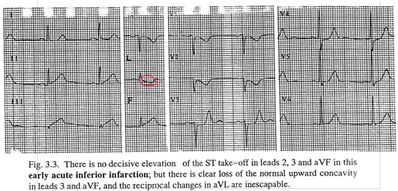
Loss of upward concavity
The earliest sign of inferior wall injury
•
•
2, 3, aVF lead ST segment straightening
•
•
“True” posterior wall infarction; reciprocal change >> indicative change
Loss of upward concavity
inferior wall injury에서 가장 먼저 발생하는 sign이며, 2, 3, aVF lead의 ST segment가 straighten됩니다.
posterior wall 에만 infarction이 발생한 경우, reciprocal change가 indicative change보다 저명하게 나타나게 됩니다.
•
aVL lead
•
inferior lead clearcut indicative change (-) & significant negative ST-T displacement in aVL
•
ST-T sagging in aVL
•
disproportionate to the size of the small QRS
•
Acute inferior infarction, NOT NONSPECIFIC ST ABNORMALITY
또한 inferior wall infarction에서 주목해야할 것은 aVL lead입니다.
inferior lead에서 명확한 indicative change가 없어도, ST-T displacement가 aVL lead에서 나타날 수 있습니다.
small QRS 와 비율이 맞지 않는 ST-T sagging이 나타나게 됩니다.
이는 acute infarction을 시사하는 소견으로, NSST가 아니라는 점을 명심해야 합니다.
Inverted U wave
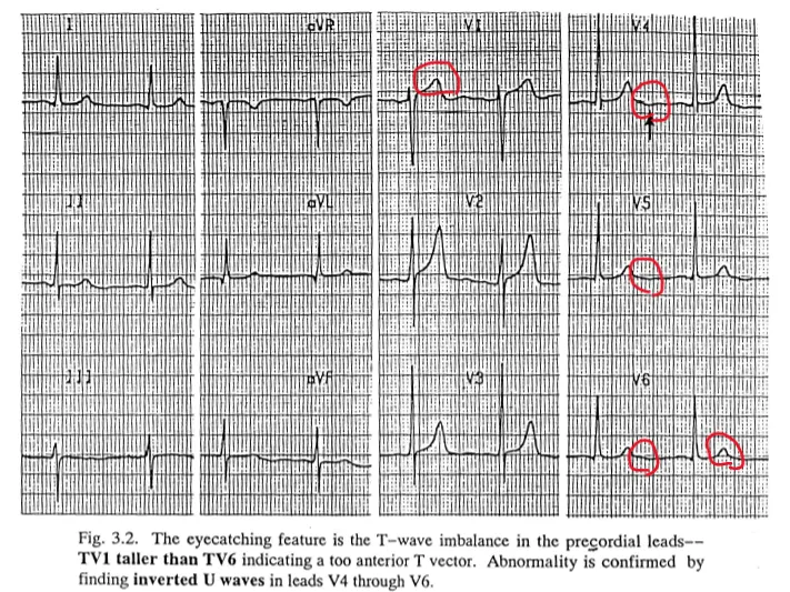
“TV1-taller-than-TV6”; T-wave imbalance in the precordial leads
Precordial lead T wave size 비교하여 V1>V6 일 때 이는
예외없는 abnormal finding은 아니나, inverted U wave 보일 시 다른 abnormal finding을 찾으려고 노력해야한다
Wellens’ warning
•
Terminal inversion of the T wave in any of the anterior precordial leads
•
SINISTER index of a proximal obstructive lesion in the Lt. anterior descending coronary artery
Mere transient ischemic attack으로 치부해서는 절대 안된다!
LAD는 angioplasty 나 bypass를 시행하여야 함
그러나 severe obstructive lesion 없이도 발생할 수는 있다
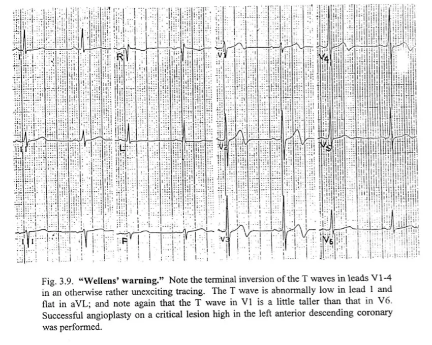
terminal inversion of the T waves in leads v1-4
Wishbone effect
•
Significant elevation of the ST take-off
•
little or no actual elevation on ST
•
T wave looks two limbs had been pulled apart, wide-based (spread-eagled) T wave prominently tall
it is important to be alert to wide-based precordial T waves
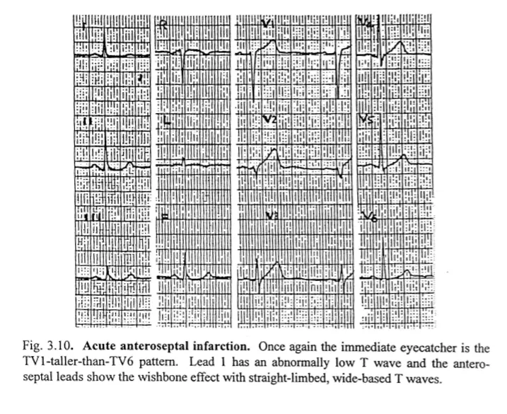
straight-limbed, wide-based T waves
Remote location
•
Posterior wall; inaccessible regions of the heart in ECG
•
Routine exam (-)
•
Indirectly by reciprocal changes in anterior leads;
•
Tall R waves in V1, V2
•
Depression of the ST segment in V1
Delayed presentation
환자들의 reluctance 혹은 Stoicism(고통을 참으려는 경향) 으로 18-24시간 가량 진단이 delay되는 경우가 있을 수 있음
R wave 소실 및 T wave inversion이 발생하나 앞서 다뤘던 전형적인 양상의 ECG들과 다를 수 있다
확진 될 때까지 disposition하면 안된다
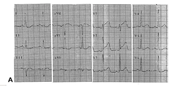
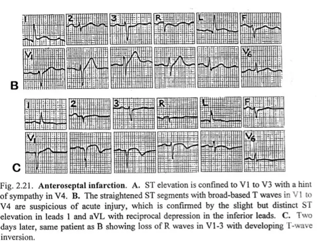
Anteroseptal infarction, remote onset case
A: 방금 막 왔을 때 찍은 EKG: V1-3 ST elevation 확인됨
B: straightening ST segment change 확인됨-> 명확하지는 않아서 놓칠 수 있음
C: 이틀 뒤 EKG: T wave inversion, ST elevation
Minor chamber involvement
•
Atrium
•
Atrial tachyarrhythmia
•
Displacement of PR segment
•
•
RV
•
V1 ST elevation, V2 ST depression Rt. sided chest wall leads (V4R)
•
RV, atrium infarction 은 inferior myocardial infarction과 동반될 수 있음을 기억해야한다.
항상 고려해야 함
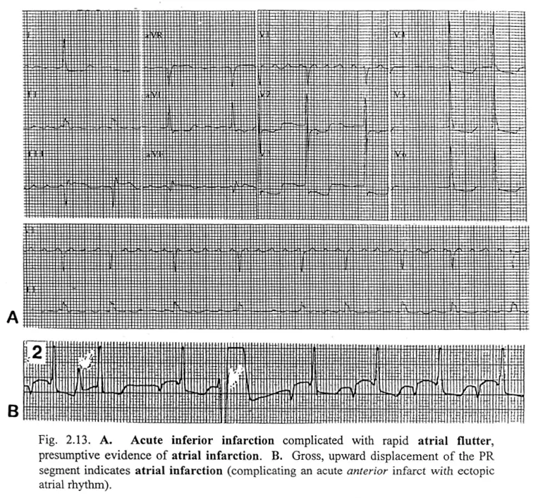
A. A. flutter ->atrial infarction 동반된 inferior infarction
B. PR segment displacement -> atrial infarction
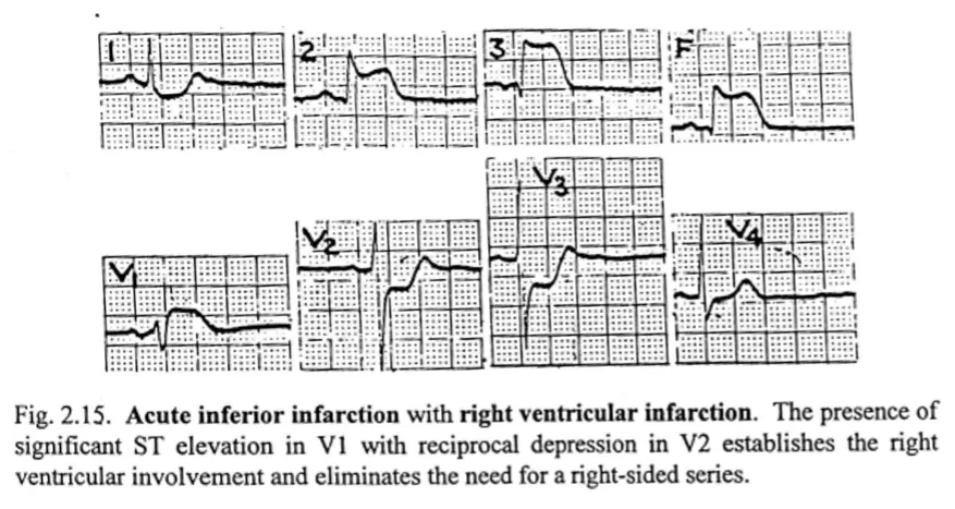
V1 STE, V2 STD -> right sided series 꼭 찍어봐야한다 (RV infarction)
