aVR STE
de Winter sign
Wellens’ syndrome
LBBB with STE
Multivessel disease
Diagonal branch occlusion
LCX artery (posterior wall MI)
A. aVR ST elevation
A1. aVR STE 원인
•
Proximal LAD occlusion
•
Severe triple vessel disease
•
Diffuse subendocardial ischemia (due to O2 supply/ demand mismatch)
A2. 기전
•
aVR은 I, II, aVL 및 V4-6과 전기적으로 반대에 위치
⇒ 나머지 lead에서의 depression은 aVR에서의 reciprocal ST elevation을 유발
⇒ Diffuse한 subendocardial ischemia에서 aVR ST elevation이 나타남
•
aVR은 RVOT와 심실 중격의 기저부를 포함하는 심장의 우측 상방 부분을 직접 기록
⇒ 이 부위의 경색이 있는 경우 aVR의 ST elevation을 유발할 수 있음.
A3. widespread ST depression + MI의 증상이 있을때
•
STE in aVR ≥1mm 는 proximal LAD/LMCA 의 occlusion이나 severe 3-vessel disease 를 시사합니다.
proximal LAD occlusion과 LMCA의 occlusion을
감별할 시에는 aVR lead와 V1 lead의 ST를 비교해야합니다. aVR lead 의 ST elevation이 V1 lead에서보다
높을때 LMCA의 occlusion을 시사함.
•
LVH에서도 St depression이 나타날 수 있는데, LV strain이라고도 합니다. 이는 비후된 근육의 repolarization의 변화나 subendocardial ischemia에 의해 유발될 수 있습니다. LVH에 의한 ST depression과 다른 원인을 감별하기 위해서는 S, R wave가 criteria를 만족하는지, asymmetric한 ST depression인지를 관찰함으로써 판단할 수 있음.
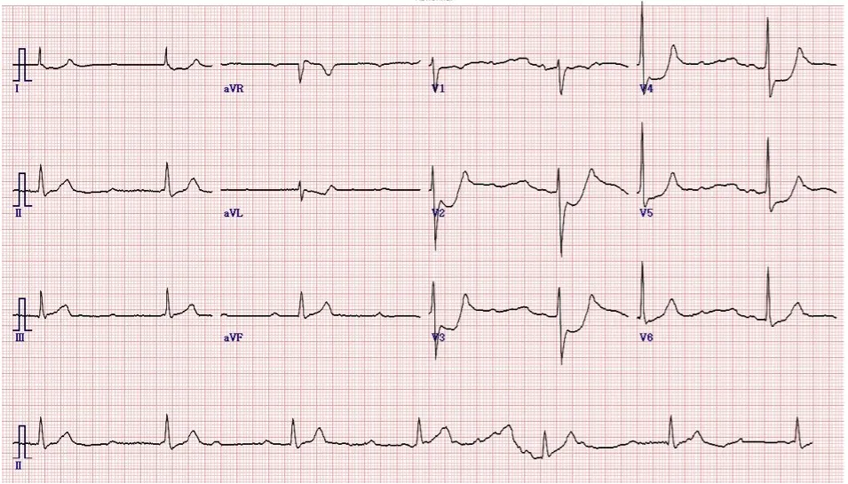
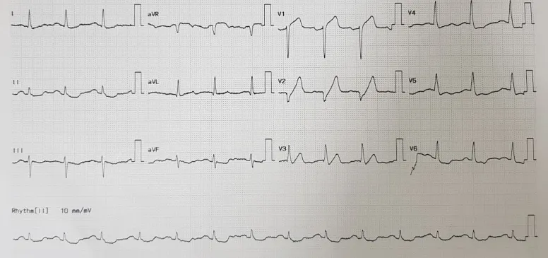
A4. 실제환자사례
•
M/76, HTN, 고지혈증 Hx. 2달 전 시작된 흉통과 등으로 방사되는 통증, 호흡곤란
•
심전도
intraventricular conduction delay
Cornell criteria SV3+R aVL이 28mm 이상을 만족하는 LVH 소견 보였습니다.
Lead I, II, III, aVF와 ,V4-6 에서 widespread한 ST depression과 aVR에서 ST elevation
•
troponin I : 0.25로 상승
•
CAG 결과 : distal RCA, proximal RCA, proximal LCX, mLAD에서 narrowing 관찰되어 triple vessel disease임을 확인
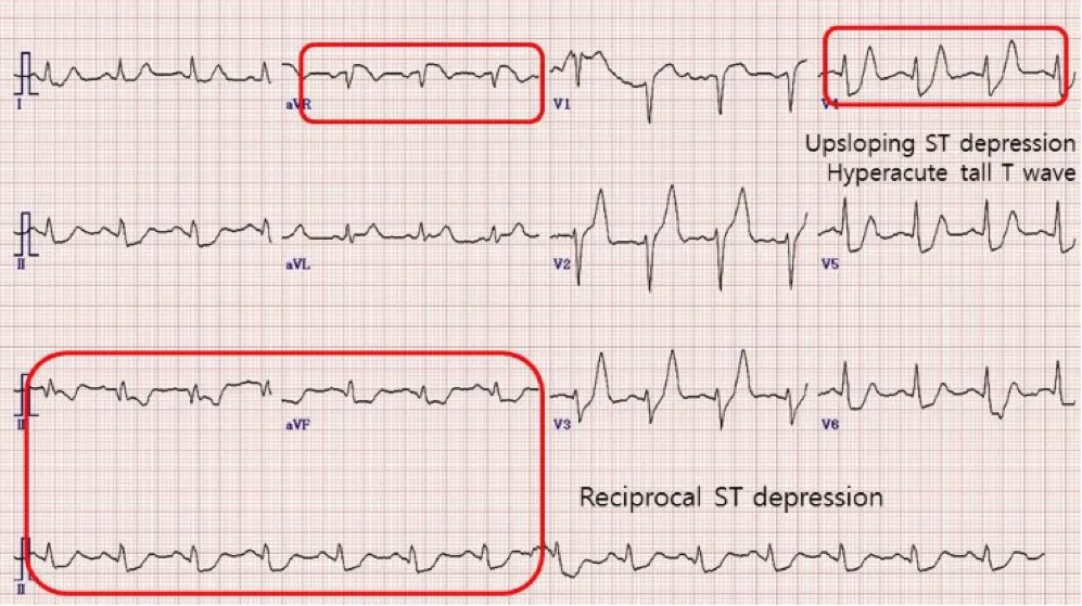
B. De Winter sign : Acute proximal LAD total occlusion
• 전벽 STEMI 의 2%에서 나타남
• pLAD의 total occlusion 의미
• Collateral circulation
• Wraparound LAD (50%)
• One vessel dz (67%)
< ECG 특징 >
• Upsloping STD 1~3 mm at J-point in V1~V6
• Hyperacute tall T wave
• aVR ST elevation
• Reciprocal ST depression in Inf. Leads
• Wellens와 감별 : Wellens는 biphasic 이거나 V2, V3에서 inverted T를 보인다
(만성 high grade LAD stenosis)
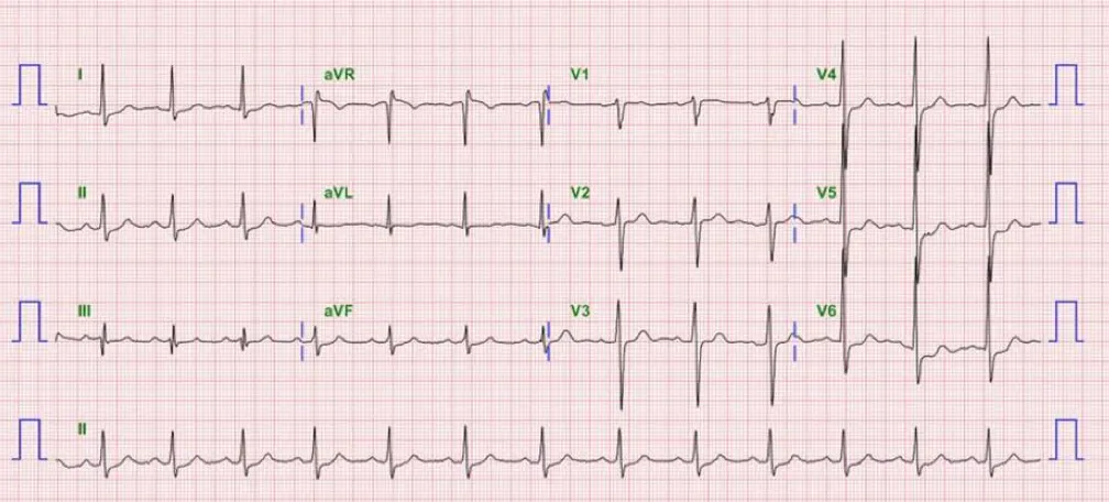
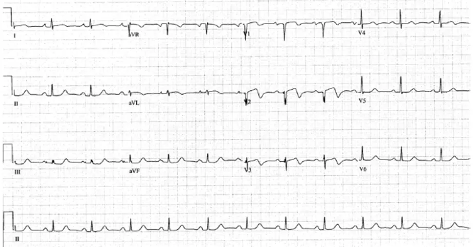
C. Wellens' Syndrome : Prox LAD 의 치명적 폐쇄가능성
•
Wallen's syndrome의 진단기준은 During pain free period
◦
Progressive, deep T wave inversion in V2, V3(V4)
◦
ST-T angle: 60-90˚
◦
T inversion: symmetric(biphasic)
◦
Little or no ST elevation
◦
No loss of R progression
During pain
•
Positive T wave
•
ST elevation
During pain free period
•
T wave inversion
•
Little or no STE
T wave inversion
•
Spontaneous reperfusion
Patient
•
Longstanding angina
•
Collateral circulation
1. Prior history of chest pain
2. 흉통있을때 : 정상 EKG or mild STE or STD or terminal T wave inversion in V1 and V2
3. 심근효소는 정상이거나 약간증가
4. No pathologic precordial Q waves
5. No poor R : 살아있는 심근많다
6. 흉통없을때 : Deeply(>2mm) inverted or biphasic T in V2 and V 3, possibly V1, V4, V5 and/or V6
•
Unstable angina(14-18%)
•
Specific ECG sign - pain free period
•
Troponin: no or little elevation
•
Critical stenosis of proximal LAD
•
Impending extensive ant. MI
•
Need emergency PCI
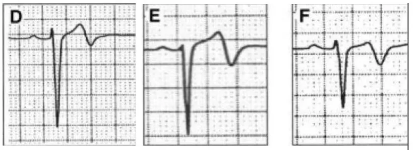
C1. 두 가지 종류
(1) Symmetrical, deep T wave inversion in V2, V3 (V4): ST-T angle: 60-90˚ - 76%
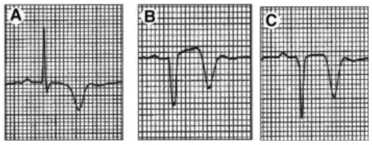
(2) Biphasic T wave inversion in V2, V3 - 24%
•
Little or no ST elevation
• No loss of R progression

C2. 실제환자
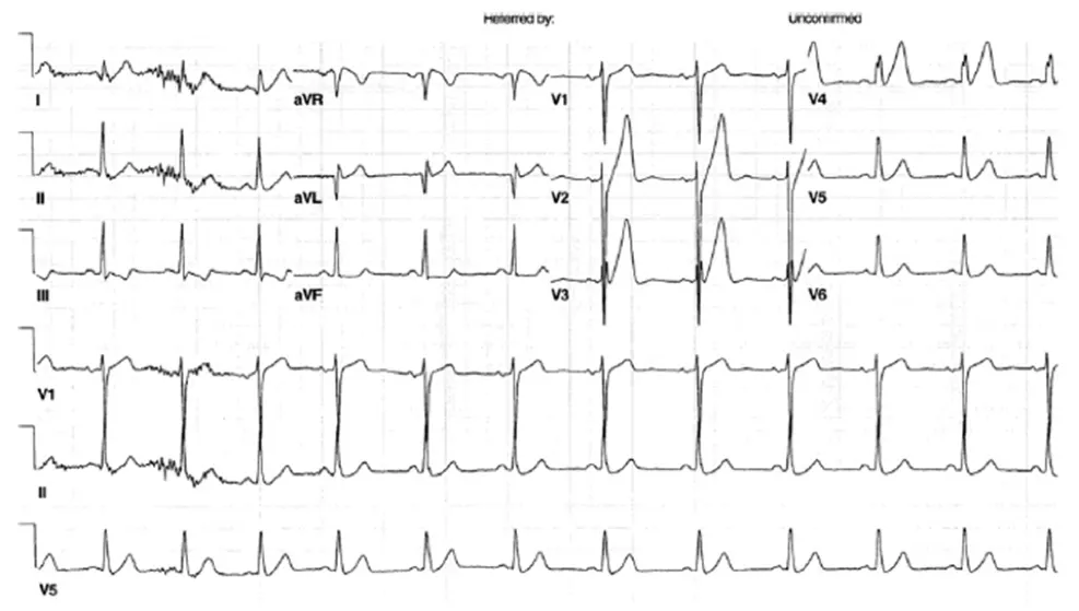
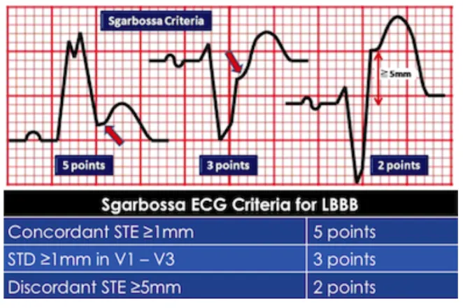
D. LBBB 에서 Sgarbossa's criteria 만족
D1. Sgarbossa’s criteria
(1) LBBB 또는 심실기원 박동이 있을 때 심근 경색식별 위한 심전도 소견들을 모아놓은 것
(2) LBBB가 있는 경우 심근경색을 감지하기 어려워서 고안된 것.
(3) 3점 이상일 경우 STEMI에 대해서 90%의 특이도(단 민감도는 36%로 낮음)
•
하나 이상의 유도에서 concordant STE - 5점
•
V1-V3 중 하나 이상의 유도에서 concordant STD - 3점
•
Negative QRS (Discordant) 유도에서 STE ≥5 mm - 2점
•
V1-V4 유도에서 비례적으로 과도한 discordant STE
ST/S ratio of equal to or more than 0.20 and at least 2 mm of STE -2점
E. Multivelssel disease
Diffuse subendocardial ischemia
•
Circumferential subendocardial ischemia
•
Lt main dz/Multivessel dz
•
Cardiogenic shock/VF
•
High mortality
•
Emergency PCI indication
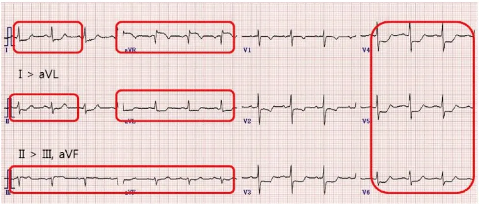
ECG findings
Diffuse ST depression(>6 leads)
•
Ant. Leads: V3-6(V4)
•
Lat. Leads: I >aVL
•
Inf. Leads: II >aVF, III
ST elevation
•
aVR(>0.5mm), V1
•
Lt main dz: aVR>V1
Direction of T wave
•
V4-V6
•
Negative T: poor prognosis
•
Positive T: more favorable outcome
Diffuse ST depression( > 6 leads)
• Ant. Leads: V3-6(V4)
• Lat. Leads: I > aVL
• Inf. Leads: II > aVF, III ST elevation
• aVR ( > 0.5 mm),V1 : Multivessel infarction인 경우 ST Vector는 RUQ로….
• High mortality
• Emergency PCI indication
F. Lt Circumflex artery
Posterolateral wall MI
•
ST depression in V1V3
•
Downsloping ST depression
•
Positive T wave
•
R/S ratio >0.5 in V1, V2(Q equivalent)
•
Left dominant: STE(II, III, aVF/V5, V6)
•
Non dominant: only STD(V1V3)